

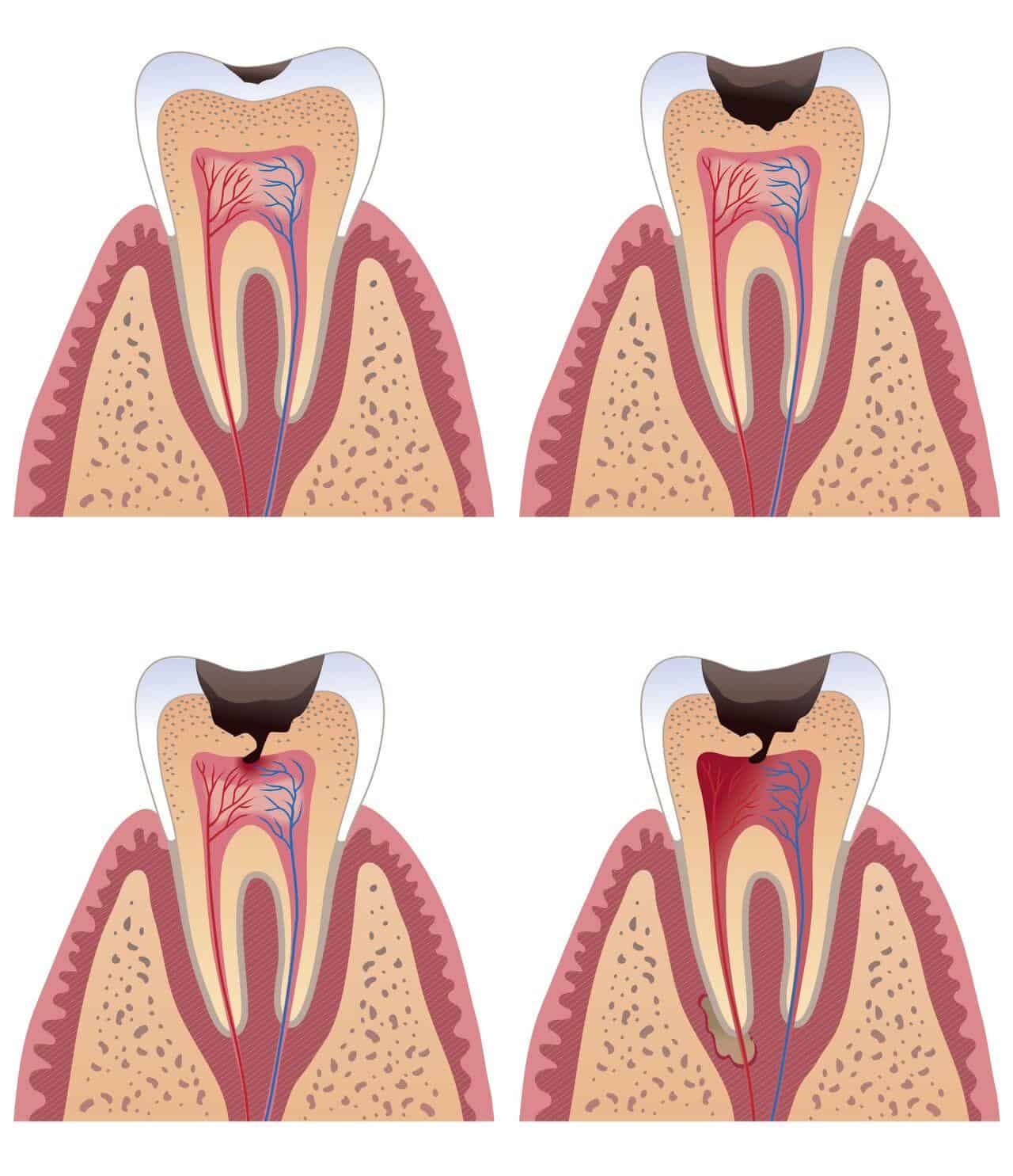
X-ray illustration of teeth
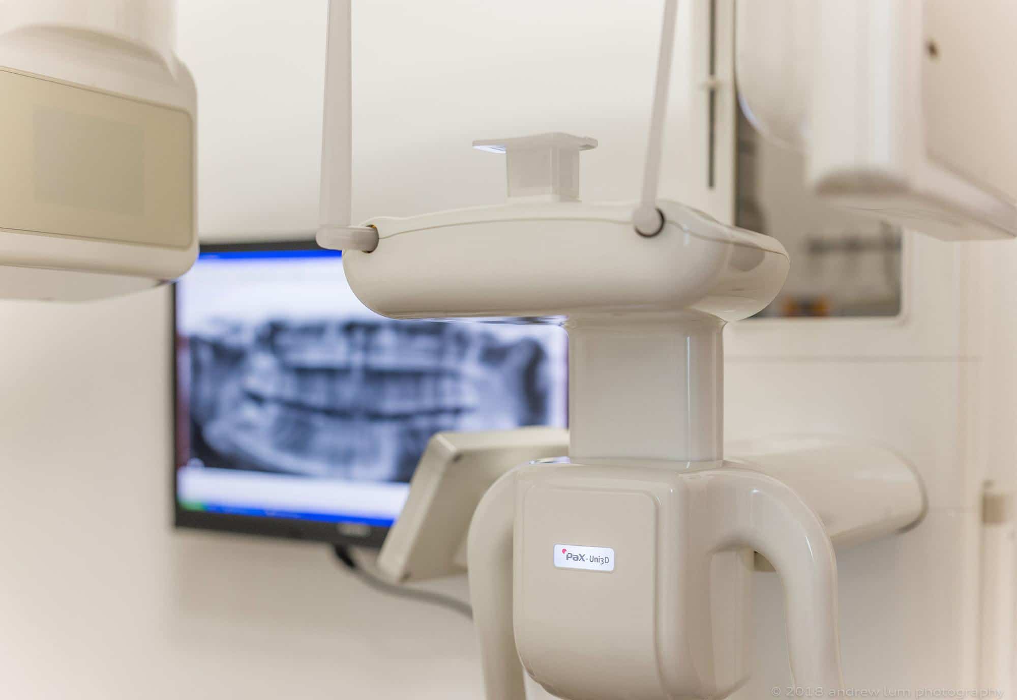
Digital Dental Imaging
Elite Dental uses only digital radiology systems. The digital radiology images or x-ray images are “developed” instantly with a scanner.
Digital dental x-rays require only about 50% of the radiation dose used for traditional film x-rays. The radiation exposure is very minimal. We are aware that some of you may have the belief that dental x-rays are potentially harmful and can even cause an increased risk of cancers such as thyroid cancer.
We have created the table below to help dispel some of the myths regarding the perceived “ high radiation” dosages of dental x-rays.
| Procedure/source of exposure | Approximate radiation dose | Comparable to natural environmental radiation for: | *Additional lifetime risk of fatal cancer |
|---|---|---|---|
| CT Head Scan | 2 mSv | 8 months | Very Low |
| Mammography | 0.4 mSv | 7 weeks | Very Low |
| Chest X-ray | 0.1 mSv | 10 day | Negligible |
| Coast to coast flight in a commercial airplane | 0.03 mSv | ||
| 2 bitewing + 2 periapical dental x-rays | 0.008 mSv | 1 day | Negligible |
| Panoramic X-ray | 0.007 mSv | 1 day | Negligible |
According to the National Council on Radiation Protection and Measurements, the average person receives an effective dose of 3 millisieverts (mSv) per year from naturally occurring radioactive materials and cosmic radiation from outer space. Naturally occurring radioactive materials can be found in the air that we breathe in and the food that we eat. Cosmic radiation is that which we receive from the sun in the form of UVA/UVB wavelengths.
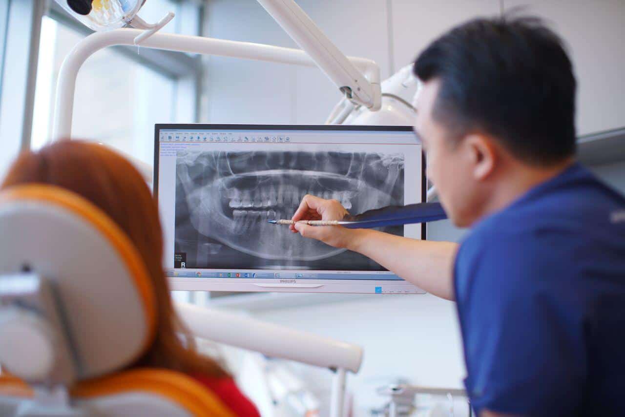
Why do I need to take dental X-rays?
X-rays are taken to detect underlying problems that cannot be seen just by looking into the mouth with the naked eye; for example, impacted wisdom teeth, bone loss due to gum disease, possible decay in between teeth and under old fillings, cysts and abscesses in the jaw etc.
It is our standard of care for new patients to undergo dental X-rays for a clear picture of your dental health, especially if you do not have any X-rays from your previous dentist. It is easy to miss problems if dental X-rays are not taken, especially when there are no complaints of pain or discomfort.
Some patients may even accuse the dentist of not being thorough in the check up if dental X-rays are not suggested. Early detection and treatment helps you to avoid emergencies and expensive dental work later on.
You may choose to skip x-rays if you are pregnant, or if you have had dental x-rays taken recently at another dental practice. Please provide any recent dental x-rays by email ahead of your appointment.
These are some of the different dental X-rays that your dentist may order:


